

Penis Envy Spores: Identification Under the Microscope
What do Penis Envy spores look like under a microscope? Quick Answer
Penis Envy spores are the microscopic reproductive cells of Psilocybe cubensis — the species to which this cultivated strain belongs. Under transmitted brightfield light, they appear as dark purplish-brown, ellipsoid to subellipsoid structures measuring approximately 11–17 µm in length and 7–12 µm in width. Their thick, smooth spore wall gives them a robust appearance, and deep pigmentation makes them identifiable at 400×. A defining feature — the apical germ pore — becomes visible at 1000× oil immersion. UK hobbyist microscopists use spore syringes to prepare wet mounts for observational study. Explore Fungi Myco's Penis Envy spore syringes, prepared specifically for microscopy research.
Taxonomy, Classification, and Strain Identity of Penis Envy Spores
There are conflicting versions as to the origins of the Penis Envy mushroom strain but we do know that they trace back to the 1970s. Its name comes from the unusual shape of its mature fruiting bodies, thick-stemmed with a small, rounded cap, characteristics that also correspond to the way its spores develop and distribute.
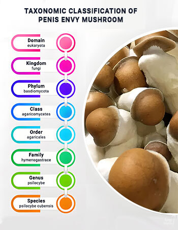
"Penis Envy" is not a species. It is a cultivated strain, a product of selective human intervention, and it holds no independent entry in any mycological registry. The accepted binomial is Psilocybe cubensis (Earle/Singer) 1948, as recorded by Index Fungorum [1] and confirmed in the NCBI Taxonomy Browser [2].
The full classification places this species within:
This distinction matters enormously for anyone building a reference collection in the UK: when you study a PE spore slide, you are observing Psilocybe cubensis traits, not a unique variety.
Understanding strain identity also means understanding what microscopy can and cannot confirm. No morphological feature visible under a compound microscope uniquely identifies a cultivated strain. Penis Envy Uncut, Albino PE, DC Melmac, and standard Psilocybe Cubensis varieties all share the same spore blueprint at the structural level — ellipsoid shape, thick smooth wall, deep pigmentation, and a single apical germ pore. What differentiates them at the cultivation level — phenotypic traits such as fruiting body shape, cap morphology, and veil behaviour — is entirely absent from a spore slide. UK mycologists working from spore-only reference collections must therefore ground their records in species-level taxonomy, not strain labelling, if scientific rigour is the goal.
The practical implication for UK researchers is that a well-maintained reference log should record species name, strain provenance, source, preparation date, and microscopic observations — but should not assert strain identity based on spore appearance alone. The Natural History Museum's Fungi department exemplifies the standard of taxonomic rigour that serious amateur mycologists in the UK can aspire to replicate at the observational level [3].
Magnification, Slide Preparation, and the Limits of Strain-Level Identification
Unlike many other cubensis strains, Penis Envy produces fewer spore deposits but with a much denser pigmentation and thicker spore walls. These qualities make its spore prints especially distinctive and valuable for close study.
Getting a clean, informative PE slide is half the identification work. 40–100×is your orientation zone: locate spore clusters, assess overall distribution density, and scan for contamination. With PE, expect a sparser field than you might see with open-veiled strains — the closed-veil tendency of PE fruiting bodies directly limits spore release and collection. 400× is where identification begins in earnest: ellipsoid shape becomes clear, pigmentation is vivid, and wall integrity is assessable. This is the working magnification for most routine Psilocybe cubensis morphology work and the level at which UK researchers can confidently record shape and colour observations. 1000× oil immersion is required for the apical germ pore — apply a small drop of immersion oil to the coverslip and rotate the oil-immersion objective into place carefully.

For slide preparation from a spore syringe: shake firmly to resuspend settled spores, deposit 1–3 µL onto a clean glass slide, and apply a coverslip gently. For PE specifically, allow the syringe to sit tip-down for several minutes before sampling, since lower spore density means more spores settle toward the tip. Wet mounts suit immediate study; air-dried preparations suit long-term reference collections. Phase contrast or darkfield optics enhance contrast without chemical staining. Melzer's reagent can test for amyloid wall reactions — a useful supplementary data point for thorough records.
Now for an important limit: you cannot reliably distinguish a Penis Envy spore slide from a Golden Teacher or B+ slide by microscopy alone. All Psilocybe cubensis strains share the same fundamental spore blueprint. A peer-reviewed PMC authentication study on Psilocybe specimens makes this clear: even expert morphological analysis produces misidentification errors, and DNA-based methods are required for definitive strain-level determination. The most practically observable difference for UK researchers is deposit density — PE and its variants consistently produce lower spore counts per slide than Golden Teacher or B+. Albino PE spores also appear measurably paler under 400× brightfield.
The UK Legal Framework for Studying Penis Envy Spores
The legal position for UK microscopists is clearer than many assume — and it is worth stating plainly.
Psilocybe cubensis spores do not contain psilocybin or psilocin. Those are the substances classified as Class A under the Misuse of Drugs Act 1971. Ungerminated spores — including Penis Envy Spores — contain neither compound. Possession of spores for microscopy and taxonomic study is therefore lawful in the UK under current legislation [4].

The key legal boundary sits at intent and action. The MDA 1971 controls controlled substances, and psilocybin only develops during germination and mycelial development. Spores held and used for microscopy research occupy a categorically different legal space from materials intended for cultivation. That distinction is not a loophole — it reflects the actual chemistry and the actual legislation.
UK researchers understandably want reassurance on this point before committing to a study practice. The reassurance is legitimate, but so is the responsibility to stay informed. Legislation can change, and the intent framing matters: maintaining clear, honest records of microscopy activity is simply good practice — both scientifically and from a compliance standpoint. If you have specific legal questions, consult a qualified legal professional rather than relying solely on educational content.
Fungi Myco supplies spores labelled explicitly for microscopy and taxonomic research, consistent with UK community norms and the expectations of the responsible mycology community. Our materials are prepared to a standard that UK researchers can rely on — sterile, clearly labelled, and supplied with the educational context that serious study demands. If you have questions about our research materials or want guidance on getting started, get in touch with the Fungi Myco team directly.
Studying these spores responsibly, with scientific rigour and legal awareness, is exactly what mycology should look like in the UK. The microscope is a tool for understanding — and that understanding begins with knowing where the boundaries are.
FAQs
Under transmitted brightfield light, Penis Envy spores appear as dark purplish-brown, ellipsoid to subellipsoid structures. At 400× they show a smooth, thick cell wall and deep pigmentation. At 1000× oil immersion, a single apical germ pore becomes visible at one end of the spore — the most diagnostic feature of Psilocybe cubensis.
Penis Envy spores fall within the published size range for Psilocybe cubensis: approximately 11–17 µm in length and 7–12 µm in width. Because PE is a cultivated strain rather than a distinct species, its spore dimensions overlap with other cubensis strains. Accurate measurement requires a calibrated eyepiece graticule or digital imaging software.
Both are Psilocybe cubensis strains and share the same fundamental spore morphology — ellipsoid shape, purplish-brown pigmentation, and an apical germ pore. The most practically observable difference is deposit density: PE produces significantly fewer spores per slide. Albino PE variants also show reduced pigmentation. Definitive strain-level separation by microscopy alone is not reliably achievable.
Penis Envy is known to produce partially or fully closed veils in many fruiting bodies, which limits the natural release and collection of spores compared to open-veiled strains like Golden Teacher or B+. This results in lower spore density in syringes and prints, meaning practitioners typically see fewer spores per microscopy slide and may need to sample more carefully.
Yes. Psilocybe cubensis spores do not contain psilocybin or psilocin — the substances controlled under the Misuse of Drugs Act 1971. Possession of spores for microscopy and taxonomic research is lawful in the UK. The legal boundary is reached if spores are germinated with cultivation intent, which falls outside the scope of legitimate microscopy study [4].
- Index Fungorum — Psilocybe cubensis Name Record. Available at: https://www.indexfungorum.org/Names/namesrecord.asp?RecordId=290107 (Accessed 25 July 2026).
- NCBI — NCBI Taxonomy Browser — Psilocybe cubensis. Available at: https://www.ncbi.nlm.nih.gov/Taxonomy/Browser/wwwtax.cgi?id=181762 (Accessed 25 July 2026).
- Natural History Museum — Fungi, lichen and bryophyte survey methods and ID guides. Available at: https://www.nhm.ac.uk/content/dam/nhm-www/take-part/monitor-and-encourage-nature/nature-recording-hub/fungi-lichen-and-bryophyte-survey-methods-and-id-guides-sheet.pdf (Accessed 2 July 2025).
- UK Government — Misuse of Drugs Act 1971. Available at: https://www.legislation.gov.uk/ukpga/1971/38/contents (Accessed 11 April 2026).