

Golden Teacher Spores: Identification Under the Microscope
What is Psilocybe Cubensis Golden Teacher? Quick Answer
Golden Teacher is a cultivar — an informally named strain — of Psilocybe cubensis, the species formally described by Franklin Sumner Earle in 1906 and transferred to Psilocybe by Rolf Singer in 1949. It carries no independent taxonomic status. Spores are subellipsoid, measuring 11–17 × 7–12 µm, with a thick smooth wall, dark purple-brown pigmentation, and a distinctive apical germ pore. In the UK, ungerminated spores are legal to possess for microscopy research.
Taxonomy and Cultivar Classification of Psilocybe Cubensis
Few fungal species have a taxonomic biography as tidy — and as frequently misread — as Psilocybe cubensis. The story begins in 1906, when American mycologist Franklin Sumner Earle formally described the species from a Cuban specimen, placing it in the genus Stropharia under the name Stropharia cubensis. That designation held for over four decades. Then, in 1949, Rolf Singer made the transfer that stands today: the species moved into Psilocybe, reflecting revised understanding of its morphological and chemical affinities. UK and global mycologists now cite this 1949 reclassification as the accepted taxonomic act.
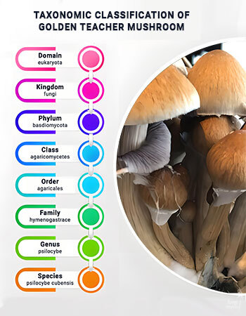
The full Linnaean hierarchy reads as follows:
Two authoritative databases maintain this record. The Index Fungorum [1] lists Psilocybe cubensis as the accepted name, while the National Library of Medicine NCBI Taxonomy Browser provides the full genomic lineage for the species [2]. Neither registry acknowledges strain-level names — a point that matters enormously.
Language matters in taxonomy, and nowhere is loose language more misleading than in the common conflation of cultivar names with species designations. A cultivar is an informally maintained line selected for particular traits: growth vigour, cap colour, or substrate preference. The name Golden Teacher belongs firmly in the cultivar category. So do names like B+, Penis Envy, and Albino A+. None appear in Index Fungorum. Treating them as species — as a notable volume of online content does — is simply incorrect.
The Golden Teacher cultivar is believed to have emerged in the 1980s, though precise provenance is difficult to verify. A peer-reviewed DNA authentication study published on PMC [3] found that misdetermination of Psilocybe specimens is common even in research contexts. For UK-based mycologists, this underscores the importance of species-level taxonomic grounding when sourcing material for microscopy study.
Spore Morphology and Macroscopic Identification of Psilocybe Cubensis
Psilocybe cubensis spores are subellipsoid: roughly elliptical with one end slightly more tapered than the other. Under transmitted light, they present as dark purple-brown, a pigmentation dense enough to be immediately visually striking at 400× magnification. Published morphometric data places spore dimensions at 11–17 × 7–12 µm — measurements consistent across cultivars, including Golden Teacher, because they reflect species-level biology rather than strain variation.
The spore wall is thick and smooth. No ornamentation. No roughening. That smoothness, combined with characteristic pigmentation, is part of the morphological fingerprint. The apical germ pore deserves particular attention: located at the apex of the spore, it is a narrow, truncated opening through which the germ tube emerges during germination. Under oil immersion at 1000×, it appears as a subtle flattening at the spore tip — confirming its presence is a critical step in species-level identification. Basidia are clavate and typically 4-spored, consistent with broader Hymenogastraceae morphology.
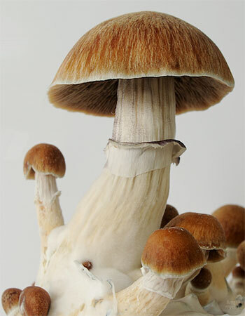
Before the microscope, there is the eye. Macroscopic features provide the first point of identification. The cap (pileus) starts convex and golden-brown with a pronounced central umbo, flattening and paling toward the margins as it matures. The surface is viscid when moist. Gills are grey when young, deepening to dense purple-black as spores mature. The stipe is fibrous and hollow, carrying a persistent membranous annulus — the remnant of the partial veil — which accumulates dark spore deposits over time.
Blue bruising is perhaps the most striking identification feature. Damage the tissue and a rapid blue-green colouration develops within seconds, resulting from the enzymatic oxidation of psilocin. Lookalike awareness is essential: Psilocybe subcubensis is morphologically similar and reliably distinguished only by spore size and microscopic features. UK academic interest in this species is grounded in taxonomy and ecology.
Microscopy Techniques for Examining Psilocybe Cubensis Spores
Good microscopy is methodical. There are no shortcuts that yield reliable data.
Begin with slide preparation. Suspend spores in sterile distilled water or isotonic saline — both media preserve morphology without inducing osmotic distortion. Transfer a small volume to a clean glass slide, apply a coverslip carefully to minimise air bubbles, and allow the preparation to settle before viewing. A drop of 3% KOH solution can be added at the coverslip edge: capillary action draws it under, and the mild alkali enhances wall visibility by reducing optical interference from cytoplasmic contents.
Start at 400×. This magnification gives you orientation — spore shape, approximate size, pigmentation, and morphological anomalies are clearly visible. Shift to 1000× oil immersion for germ pore confirmation and fine wall detail. The germ pore, at this magnification, becomes identifiable as a truncation at the spore apex — a diagnostically meaningful confirmation for UK researchers undertaking systematic study.

Accurate measurement demands micrometry. A calibrated eyepiece graticule, cross-referenced against a stage micrometer, allows you to express spore dimensions in micrometres and compare them against the published range of 11–17 × 7–12 µm. Two reagents extend the analysis. Melzer's reagent can reveal amyloid or dextrinoid reactions in spore walls, though P. cubensis spores are inamyloid — a useful negative result. Cotton blue in lactophenol stains chitin in fungal cell walls, aiding tissue differentiation when examining cystidia at higher magnification.
Spore print collection underpins all of this. Place a mature cap gill-side down on glass or aluminium foil, cover to prevent air currents, and leave for 4–12 hours. The resulting deposit — a deep purple-black print — provides the material for slide preparation. All of the above is conducted within the context of UK law: ungerminated spores contain no controlled substances and are legally possessed for research and microscopy.
UK Legal Framework and Research Context for Psilocybe Cubensis Spores
Clarity here is not optional — it is the foundation of responsible engagement with this subject.
Under the Misuse of Drugs Act 1971, psilocybin and psilocin are classified as Schedule 1 controlled substances in the UK [4]. Cultivation of Psilocybe cubensis fruiting bodies — the process by which these compounds are produced — is therefore illegal. This is unambiguous.
The legal position of ungerminated spores is equally clear, and equally important. Spores of Psilocybe cubensis contain neither psilocybin nor psilocin. They are, in their ungerminated state, not controlled under the 1971 Act. Possession, purchase, and use of spores for microscopy and taxonomic research purposes is lawful in the UK. This is the legal basis on which spore syringes and spore prints are supplied by UK-based vendors for legitimate research purposes.
The scientific interest in this species is not fringe. UK institutions including Imperial College London and King's College London have conducted clinical research into psilocybin's pharmacological and therapeutic properties. That institutional activity underscores the legitimate academic context within which spore taxonomy and morphology research sits. All content in this article is framed for research, education, and microscopy purposes. Nothing here should be construed as guidance toward cultivation.
One practical note on preservation relevant to UK researchers. Spore syringes should be stored at 2–8°C — standard refrigeration is sufficient — and kept away from UV light, which degrades spore viability over time. Spore prints fare best in cool, dry conditions in sealed packaging. Properly stored, material remains viable for microscopy for months to years. Protecting sample integrity is as much a part of good research practice as technique at the bench. The legal framework is clear, the science is legitimate, and the distinction between studying a species and producing a controlled substance is one the UK legal system recognises fully.
FAQs
Golden Teacher is a cultivar — an informally named strain — of Psilocybe cubensis. It holds no independent taxonomic status and does not appear in formal nomenclatural registries such as Index Fungorum. All its defining biological characteristics fall within the accepted species description of Psilocybe cubensis.
Psilocybe cubensis spores are subellipsoid in shape, measuring approximately 11–17 × 7–12 µm. They have a thick, smooth wall, dark purple-brown pigmentation under transmitted light, and a distinctive apical germ pore. These features are consistent across cultivars, including Golden Teacher, and are key to species-level microscopic identification.
Under a microscope at 400–1000× magnification, Golden Teacher spores appear subellipsoid with a smooth, thick, dark purple-brown wall and a visible apical germ pore. Basidia are clavate and typically 4-spored. These features are indistinguishable from other Psilocybe cubensis cultivars, as strain-level differences are not reliably identifiable by microscopy alone.
Key macroscopic identifiers include a convex golden-brown cap that flattens with age, gills that mature from grey to deep purple-black, a fibrous white stipe with a persistent membranous annulus, and a rapid blue bruising reaction when tissue is damaged. The spore print is characteristically dark purple-black.
Yes. Under the Misuse of Drugs Act 1971, ungerminated Psilocybe cubensis spores are legal to possess in the UK because they contain neither psilocybin nor psilocin. Golden Teacher spore syringes and prints are legally supplied for microscopy and taxonomic research. Cultivation of fruiting bodies, however, remains illegal.
- Index Fungorum — Psilocybe cubensis Name Record. Available at: https://www.indexfungorum.org/Names/namesrecord.asp?RecordId=290107 (Accessed 25 July 2026).
- NCBI — NCBI Taxonomy Browser — Psilocybe cubensis. Available at: https://www.ncbi.nlm.nih.gov/Taxonomy/Browser/wwwtax.cgi?id=181762 (Accessed 25 July 2026).
- PMC / National Institutes of Health — DNA Authentication and Psilocybe Misdetermination Study. Available at: https://pmc.ncbi.nlm.nih.gov/articles/PMC9764976/ (Accessed 26 July 2026).
- UK Government — Misuse of Drugs Act 1971. Available at: https://www.legislation.gov.uk/ukpga/1971/38/contents (Accessed 11 April 2026).